Mapping the Brain with Dr. Ashley VanMeter: A Day at the Center for Functional and Molecular Imaging
- Marshall Bailly
- Jul 11, 2025
- 2 min read

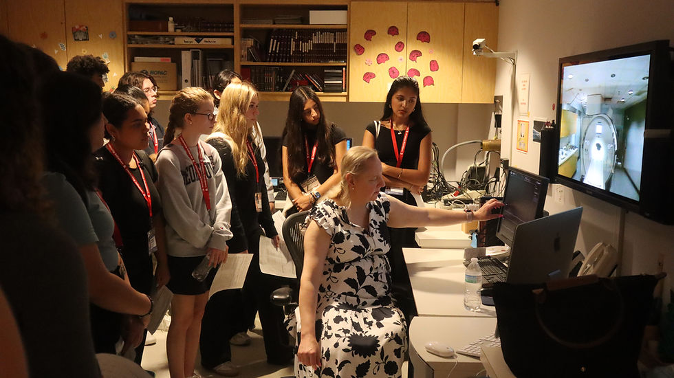
On Thursday, interns in the Advanced Medical Neuroscience Internship had the extraordinary opportunity to work with Dr. Ashley VanMeter, Director of Georgetown University’s Center for Functional and Molecular Imaging (CFMI) and a leading expert in the field of neuroimaging. Dr. VanMeter provided interns with a comprehensive look into how we study the brain using magnetic resonance imaging (MRI), offering insights drawn from her extensive research and real-world applications of advanced imaging tools.
The session began with an introduction to MRI, a non-invasive technique that uses magnetic fields and radio waves to generate detailed images of internal structures, including the brain. Dr. VanMeter explained the differences between structural MRI, which shows the anatomy of the brain, and functional MRI (fMRI), which helps researchers visualize brain activity.
Interns were especially interested in how fMRI works. This imaging method measures brain activity by tracking changes in blood flow. The idea is simple: when a specific brain region is more active, it uses more oxygen, and the body responds by increasing blood flow to that area. fMRI detects these changes using a process called BOLD imaging, which stands for Blood-Oxygen-Level-Dependent imaging. This allows researchers to observe which parts of the brain are active when a person performs various tasks, such as reading, solving problems, or processing emotions.
Dr. VanMeter shared how this tool has transformed neuroscience. One highlight of her career was co-authoring the first published study to use fMRI in investigating dyslexia. This groundbreaking research demonstrated how brain function differs in individuals with dyslexia, providing a new perspective on the condition and informing more effective intervention strategies.

She also introduced interns to other key imaging techniques such as diffusion tensor imaging (DTI), which maps the brain’s white matter pathways, and MR spectroscopy, which analyzes the brain’s chemical composition. These tools have supported her research on topics such as autism, child brain development, and how the prefrontal cortex and reward systems develop in adolescence, particularly in relation to early alcohol use.
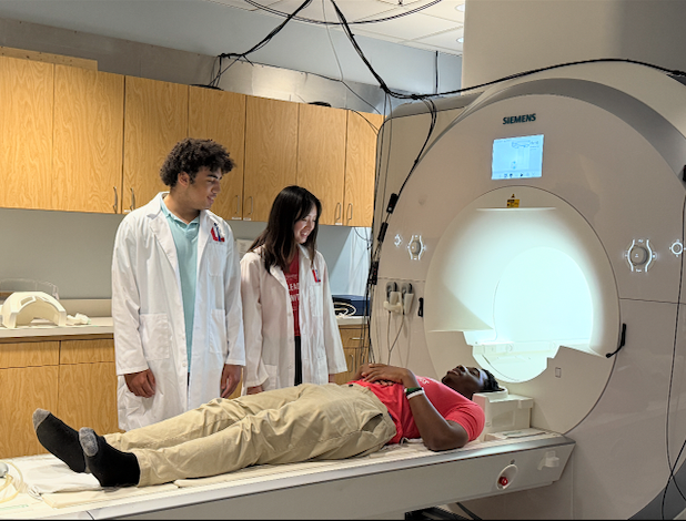
A highlight of the day was the tour of Georgetown’s dedicated 3T MRI facility. Interns learned how scientists design experiments, collect real-time data on brain activity, and analyze results using specialized software. Dr. VanMeter also discussed the role of mock scanners in preparing participants for actual scans, especially in pediatric studies, where familiarity with the equipment can improve data quality.
The session gave students a rare and valuable look at the intersection of neuroscience, technology, and research design. Dr. VanMeter's work made it clear how these tools allow scientists to ask and answer complex questions about the human brain, how it works, how it changes, and how we can support its healthy development.
By the end of the day, interns left with a richer understanding of neuroimaging and a new appreciation for the level of detail, precision, and creativity that goes into studying the brain. Dr. VanMeter’s presentation showed how far the field has come and how much potential lies ahead for the next generation of neuroscientists.


Comments